Figure 2.

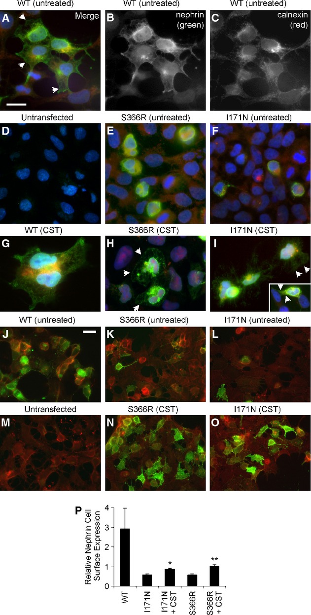
Nephrin expression and localization (immunofluorescence microscopy, Z-stack images). 293T cells were transfected with nephrin WT or the S366R and I171N mutants, as indicated. (A–F) After 48 h, cells were fixed, permeabilized and stained with antibody to the nephrin cytoplasmic domain (green) and antibody to calnexin (red). Nuclei were stained with Hoechst (blue). Calnexin staining (A and C) is generally perinuclear. Staining of nephrin WT (A and B) is in the plasma membrane (arrowheads), and there is also some perinuclear staining, which colocalizes with calnexin (A; yellow-orange staining). (D) Staining of untransfected cells with antibody to the nephrin cytoplasmic domain was negative. (E and F) In contrast to nephrin WT, the S366R and I171N nephrin mutants mainly showed perinuclear staining, and there was costaining with calnexin. (G–I) Cells were incubated with castanospermine (CST; 1 mmol/L, 18 h). Cells were fixed, permeabilized, and stained with antibodies as in A–F. Castanospermine had no significant effect on nephrin WT (G), but increased the amount of nephrin S366R and I171N appearing in the plasma membrane (arrows). (J–L, N–O, and P) At 48 h after transfection, nonpermeabilized cells were fixed and stained with antibody to the nephrin extracellular domain (green). Then, cells were permeabilized and incubated with rhodamine phalloidin (red) to visualize F-actin. Some cells (N and O) were first treated with castanospermine, as above. Nephrin WT is expressed on the cell surface (J), whereas surface expression of nephrin S366R and I171N is detected in only a few cells (K and L). Castanospermine increased expression of nephrin S366R and I171N on the cell surface (N and O). (M) Staining of untransfected cells with antibody to the nephrin extracellular domain was negative. Panels A–I and J–O are at similar magnifications; the bars = 15 μm. (P) Quantification of fluorescence intensity (green, which reflects nephrin plasma membrane expression, and red, reflecting total F-actin; see Materials). Nephrin expression was normalized for F-actin content. Castanospermine enhanced plasma membrane expression of the nephrin mutants. *P < 0.0002, **P < 0.0001 versus untreated, N = 15.
