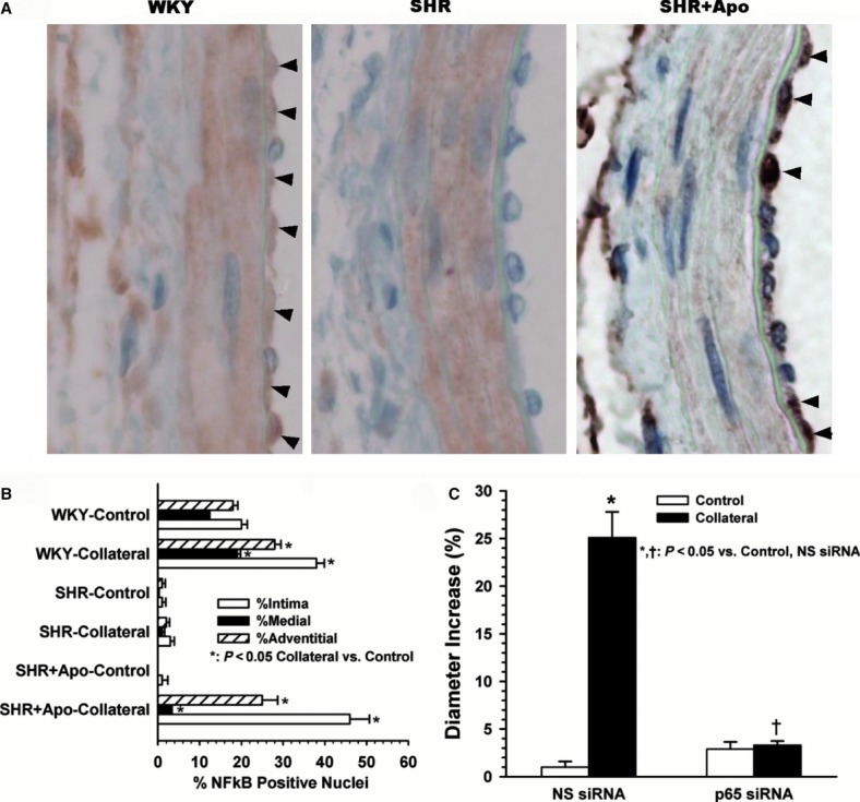
Figure 5.

Nuclear localization of NF-κB during successful and impaired collateral growth. (A) Representative images showing NF-κB immunoreactivity (brown) in collateral arteries 3 days after arterial ligation in WKY, SHR, and apocynin pretreated SHR (SHR+Apo). Nuclear localization is apparent especially within the intima of WKY and SHR+Apo, as indicated by arrows, but not in SHR. (B) Analysis of the percentage of cells with immunoreactivity in each wall layer indicates a statistical increase in all wall layers of collaterals from WKY and SHR+Apo relative to same animal controls, (n ≥ 3, *P ≤ 0.001). (C) Inhibition of p65 expression suppressed collateral growth. Paired comparisons of control and collateral arteries before and 7 days after arterial ligation demonstrated significant collateral enlargement in WKY administered a control nonsense siRNA (*P ≤ 0.001, n = 4) but not in WKY pretreated with siRNA to p65 (†P ≤ 0.001 vs. nonsense siRNA collateral, n = 3). No effect of p65 siRNA was observed on the diameters of control arteries.
