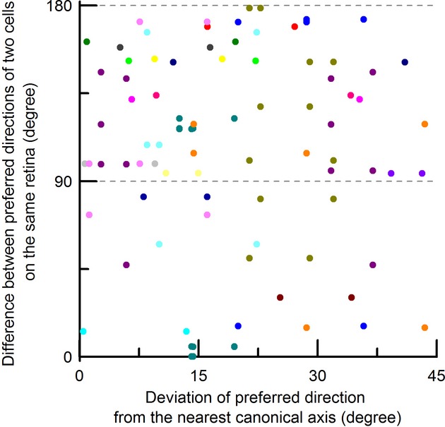
Figure 9.

The preferred directions of DSGCs on the same retina at around the time of eye opening are not aligned orthogonally or in opposite directions. The difference between preferred directions of two DSGCs on the same retina was plotted against the deviation of preferred direction of one of the DSGCs from its nearest canonical axis. The lower and upper dashed gray lines indicate the perfect orthogonal and opposite differences, respectively. Different colors are cells recorded from different retinas. The fact that most differences of preferred directions between two cells on the same retina were not distributed along two gray dashed lines suggests that the dispersion of the preferred direction observed in Figure 2A is not caused by handling and placing the retina in the recording chamber.
