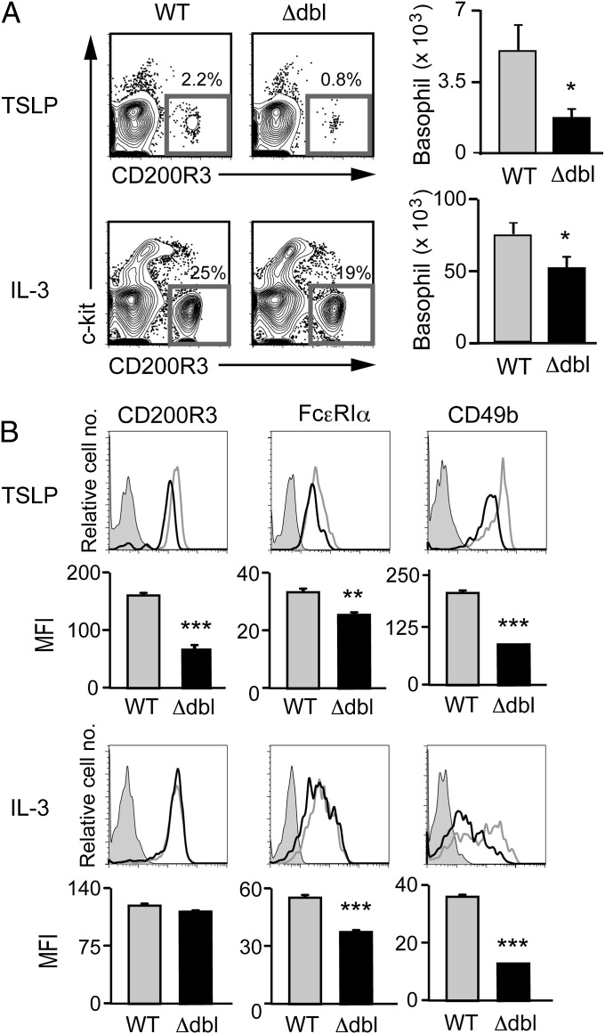
Fig. 3.
ΔdblGATA bone marrow cells show impaired generation of basophils when cultured ex vivo with TSLP or IL-3. Bone marrow cells isolated from WT or Δdbl BALB/c mice were cultured ex vivo with TSLP or IL-3 for 5 d. (A) Basophils were identified as FSClowSSClowCD200R3+c-kit− cells as shown at Left, and the number of basophils in each group is summarized at Right (mean ± SEM, n = 4 each). (B) The cell surface phenotype of bone marrow-derived basophils. Representative staining profiles are shown (TSLP and IL-3 rows); gray, black, and shaded histograms indicate those of WT and Δdbl basophils and control staining with isotype-matched antibodies, respectively. MFI of each surface marker is shown (MFI rows) (mean ± SEM, n = 4 each). Data shown are representative of at least two independent experiments. *P < 0.05; **P < 0.01; ***P < 0.001.

