Fig. 1.
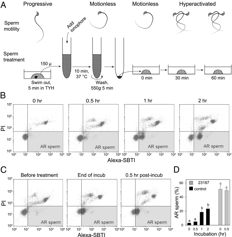
Behavior of mouse spermatozoa before and after Ca2+ -ionophore treatment. (A) Diagram showing sperm behavior and treatment. Spermatozoa were incubated in TYH for 5 min before exposure to 20 µM Ca2+ ionophore A2187. After 10 min treatment with ionophore, spermatozoa were washed and resuspended in fresh TYH. They became motionless on ionophore treatment. After washing by centrifugation, most spermatozoa showed hyperactivation by 30 min and remained so for few hours (Movies S5–S9). (B) Spermatozoa, incubated in TYH for 0–2 h, were stained with both Alexa-SBTI and PI to evaluate acrosomal status by cytometry, Two-dimensional plots differentiated between spermatozoa with low (live) and high (dead) PI staining (y axis) and those with low (intact) or high (acrosome-reacted) SBTI staining (x axis). The lower-right quadrant represented live, acrosome-intact spermatozoa; almost all live spermatozoa at 0 h of incubation were acrosome-intact. The upper-right quadrant represented dead, acrosome-disrupted spermatozoa. The lower right quadrant (colored) represents live, acrosome-reacted spermatozoa. Cytometric assays were repeated three times. Live, acrosome-reacted spermatozoa had green fluorescence in their acrosomal cap (Fig. S3). Nuclei of live spermatozoa remained dark. (C) Acrosomal status of spermatozoa before, at the end of ionophore treatment, and 0.5 h after washing the ionophore. (D) Proportions of live, acrosome-reacted spermatozoa in capacitating media at various times after starting incubation (black columns) and at 0 and 0.5 h after ionophore treatment (gray columns).

