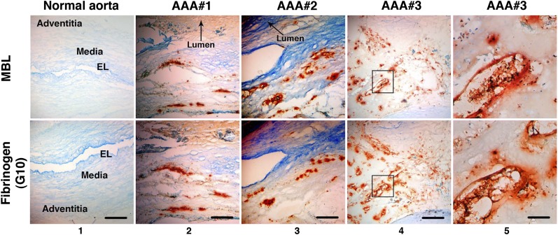
Fig. 7.
LP activation in human AAA. Immunohistochemistry performed on serial AAA sections demonstrates that MBL (orange-brown) localized to the same areas as fibrinogen (orange-brown, detected with G10 mAb) in three separate AAA tissue samples. Staining was confirmed on at least three independent sections per tissue sample. Note that the normal aortic wall architecture and layers are distorted in AAA tissues (columns 2–4) due to extensive remodeling and transmural inflammation, as evidenced by inflammatory cell infiltrates and lipid deposition (Fig. S2). Scale bar, 500 μm (columns 1–4); 100 μm (column 5). Panels in column 5 represent higher magnification of Inset in column 4. EL, elastic lamellae.

