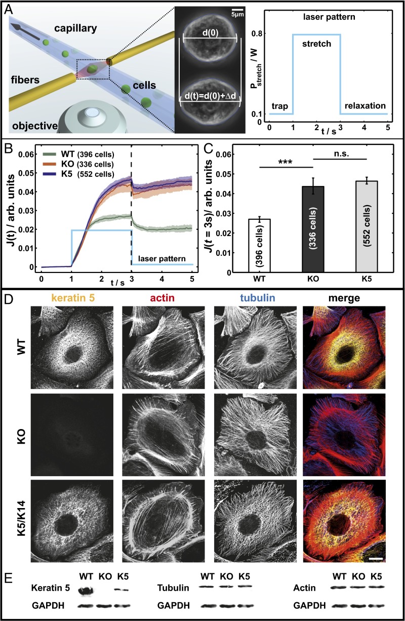
Fig. 1.
Keratins are major determinants of cell stiffness. (A) (Left) Schematic of the µOS setup showing a strongly deforming KO cell treated with LatA. (Right) Laser pattern used for all µOS measurements. (B and C) Data sets from optical stretcher measurements of keratin-free and K5/K14 reexpressing cells compared with WT cells. Characterized by creep deformation curves J(t) (B) and J (t = 3 s) (C) (***P < 0.001). (D) Immunofluorescence analysis using keratin and tubulin antibodies and phalloidin staining (actin). (E) Keratin 5, tubulin, and actin expression detected by Western blotting of total protein lysates. (Scale bar, 10 μm.)

