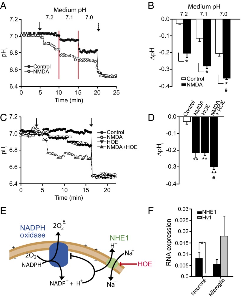
Fig. 2.
Effects of NMDA, medium pH, and Na+/H+ exchange inhibition on intracellular pH. (A) Representative single-cell BCECF traces show that both NMDA and medium acidification reduce intracellular pH (pHi), with additive effect. Arrow indicates NMDA addition, red lines delineate stepwise medium acidification, and dotted arrow shows pH 6.5 calibration. (B) Quantified aggregate results; n = 4, *P < 0.05, #P < 0.05 vs. pH 7.2. (C) Representative single-cell BCECF traces show that both NMDA and the Na+/H+ exchange inhibitor HOE (1 µM) reduce intracellular pH and have additive effects. Solid arrow indicates time of NMDA and HOE addition, dotted arrow shows pH 6.5 calibration. (D) Quantified aggregate results; n = 4, **P < 0.05 vs. control, #P < 0.05 vs. NMDA. (E) Intracellular H+ inhibits NADPH oxidase. H+ produced by NADPH oxidase and other sources may exit neurons via the Na+/H+ exchanger NHE1. HOE inhibits Na+/H+ exchange and thereby produces intracellular acidification. (F) RT-PCR assessment of cultured neurons and microglial shows that, in neurons, gene expression of NHE1 is high relative to the voltage-dependent H+ channel Hv1. (n = 4, P < 0.01).

