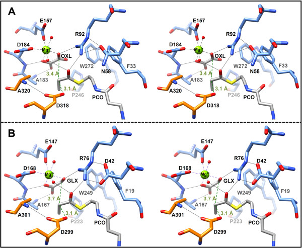
Figure 5.
Comparison of the active sites of both malyl-CoA lyases. Ligands are colored in grey. Residues of the TIM-barrel are colored in blue, whereas residues of the C-terminal lid domains of the neighboring subunits are colored in orange. Important hydrogen bonds are depicted as thin black lines. Coordination of the Mg2+ ion is shown by thick grey broken lines. Distances between the reacting α-carbon of propionyl-CoA (PCO) to the proposed active aspartate residue and oxalate (OXL) or glyoxylate (GLX) are illustrated in green. A) Stereo view of the active site of MCLC (PDB 4L80). B) Stereo view of the active site of MCLR (PDB 4L9Y).

