
Fig. 3.
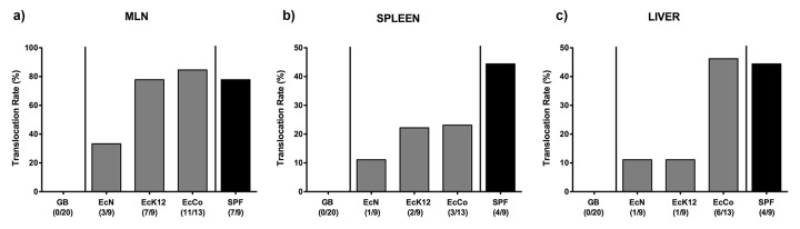
Translocation of E. coli in recolonized gnotobiotic mice following ileitis induction. Gnotobiotic mice generated by quintuple antibiotic treatment were perorally recolonized with E. coli Nissle 1917 (EcN), E. coli K12 (EcK12) or a commensal E. coli (EcCo) strain as described in methods. Four days thereafter, E. coli recolonized mice (grey bars) were perorally infected with 100 cysts of Toxoplasma gondii in order to induce acute ileitis. With conventional microbiota recolonized (SPF; black bars) and gnotobiotic (GB; white bars) animals served as positive and negative controls, respectively. Indicated are E. coli translocation rates into (a) mesenteric lymphnodes (MLN), (b) spleen, and (c) liver at day 7 following ileitis induction determined by culture of organ homogenates. Relative percentages of translocation frequencies are indicated, and absolute numbers of animals harboring the respective E. coli strain out of the total number of analyzed animals are given in parentheses. Data shown were pooled from three independent experiments.