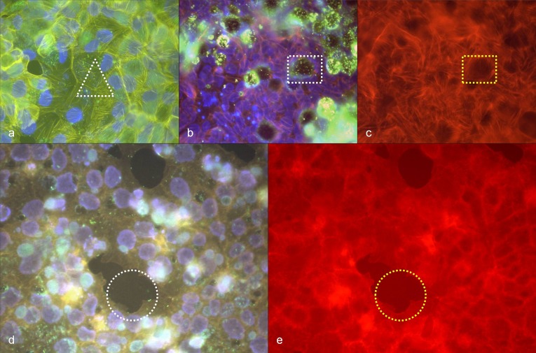
Fig. 6.
(a) C. burnetii in BGM cells, 48 h after infection, immuno-fluorescence staining (green), phalloidin-TRITC staining of actin (yellow), DAPI staining of DNA (blue), white triangle: stress fibers (yellow) without detectable association to immuno-stained C. burnetii (green); (b) C. burnetii in BGM cells, 48 h after infection, immuno-fluorescence staining (green), phalloidin-TRITC staining of actin (orange), DAPI staining of DNA (blue), white rectangle: immuno-stained C. burnetii (green) in a vacuole; (c) corresponding position to (b), phalloidin-TRITC staining of actin, yellow rectangle: absence of stress fibers around the vacuole; (d) C. burnetii in Vero E6 cells, 48 h after infection, immuno-fluorescence staining (green to turquoise in vacuoles), phalloidin-TRITC staining of actin (orange), DAPI staining of DNA (violet), white circle: detachment of infected cells; (e) corresponding position to (d), phalloidin-TRITC staining of actin, yellow circle: detachment of infected cells

