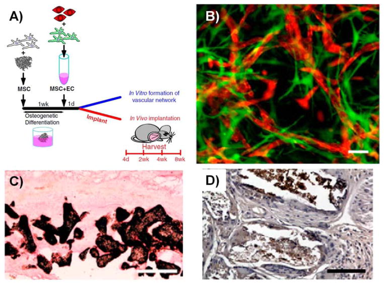
Figure 4.
(A) Tsikgou et al seeded bmMSC on PLGA scaffolds and pre-differentiated them towards the osteogenic lineage prior to embedding the scaffolds in a collagen-fibronectin hydrogel containing a bmMSC and HUVEC co-culture. (B) In vitro formation of vessel-like structures after 21 days of culture. MSC (eGFP) and HUVEC (tdTomato) were transduced with a lentivirus to fluorescently label the cells. Scale bar = 50 μm. (C) Von Kossa staining after 8 weeks of subcutaneous implantation in immunodeficient mice. Calcium deposition was observed on the pore surfaces of the implanted scaffold. Scale bar = 500 μm. (D) Osteocalcin, a late marker of osteogenesis, was also present throughout the scaffold. Scale bar = 100 μm. All panels are adapted from (118). Copyright 2010 National Academy of Sciences of the United States of America.

