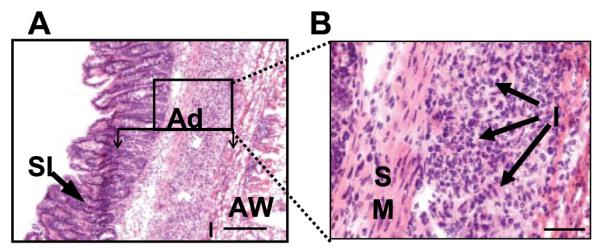
FIGURE 1.

Histologic examination of a representative surgical adhesion in which a portion of the small intestine has become attached to the abdominal wall. A, H&E staining. Original magnification, ×100. The adhesion (Ad) comprises the small intestine (SI), inflammatory infiltrate (I), and the remains of the abdominal wall (AW), which are indicated. The box indicates the field under higher magnification in B. B, The same adhesion under higher magnification. Bar, 100 μm. Original magnification, ×400. The smooth muscle (SM) of the intestine and the inflammatory infiltrate (I) are indicated. These results are representative of surgical adhesions obtained from the intestine, cecum, and intestinal wall, examined for each animal, from 10 different C57BL/6J mice.
