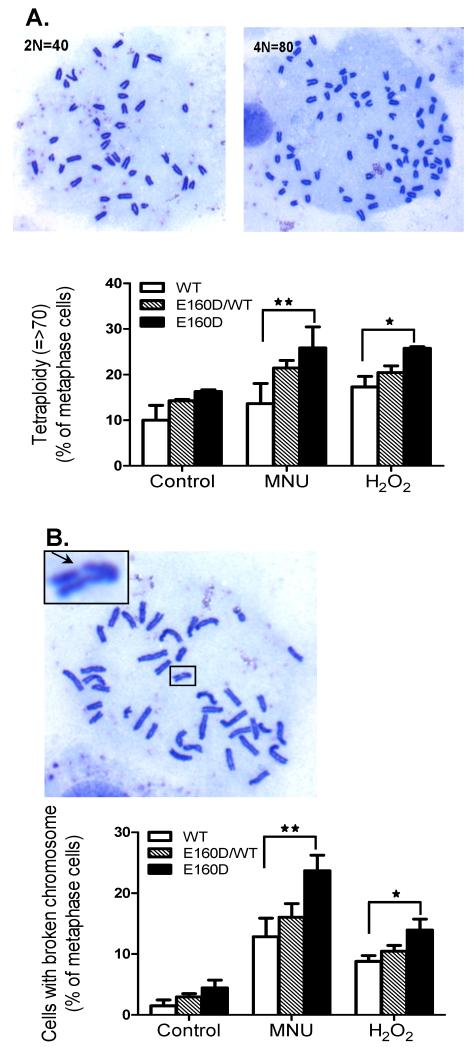
Figure 5.
MNU- or H2O2-treatment induced more chromosomal abnormalities in E160D MEF cells than in WT (A) The percentage of tetraploid and near tetraploid (n>70) chromosomes in MEF cells in WT, E160D/WT and E160D. This includes spontaneous abnormalities and those arising after chemical treatments with MNU (200 μg/ml, 1 h) or H2O2 (1 mM, 15 min). The chromosome number of each mitotic cell was counted; approximately 200 mitotic cells per cell type, per treatment were analyzed. Shown are the results of three independent experiments. *P= 0.026; **P= 0.002, Two-tailed Fisher Exact. (B) Quantification of mitotic cells (WT, E160D/WT and E160D) with broken chromosomes. The ratio was calculated by dividing the number of cells with broken chromosome by the total number of mitotic cells. *P=0.011; **P= 0.0004, Two-tailed Fisher Exact.

