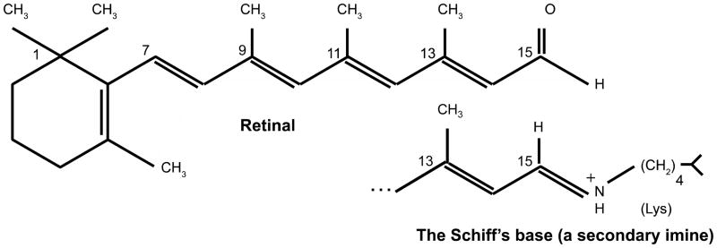
Figure 2.

In mammalian rhodopsin (MR) and in bacteriorhodopsin (BR), a lysine residue K296 (of 348 aas) in MR, and K216 (of 262 aas) in BR is covalently linked to retinal (top figure) via a Schiff’s base (bottom figure). In both cases, the Schiff’s base is in the center of TMS 7, and the retinal separates the two half channels (see text).
