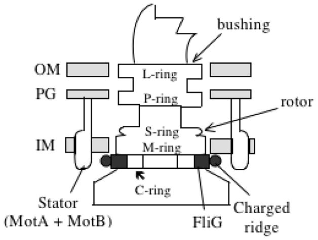
Figure 6.

Structure of the flagellar basal region and its association with the bacterial cell envelope. OM, outer membrane; PG, peptidoglycan cell wall; IM, inner membrane. The rings visible in electron micrographs are: C - cytoplasmic; M - membrane; S - soluble (periplasmic) P - peptidoglycan (PG) and L - Lipopolysaccharide-containing outer membrane (OM). The letter in the name of each ring indicates the envelope structure with which it associates.
