Figure 1.
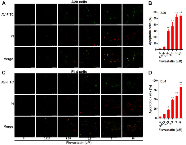
Fluvastatin induced apoptosis in lymphoma cells. Lymphoma cells were incubated with fluvastatin (0–10 μM) for 24 h. Apoptosis of lymphoma cells was analyzed by using annexin V (AV)-FITC/PI double staining and confocal laser scanning microscopy. Representative images from A20 cells (A) and EL4 cells (C) were shown (bar = 75 μm). Apoptosis levels were evaluated by the ratio of apoptotic cells (AV-FITV alone positive and AV-FITC/PI double positive cells) and total cells for A20 cells (B) and EL4 cells (D). Data are presented as mean ± SEM of three separate experiments, *p < 0.05, **p < 0.01 versus resting cells.

