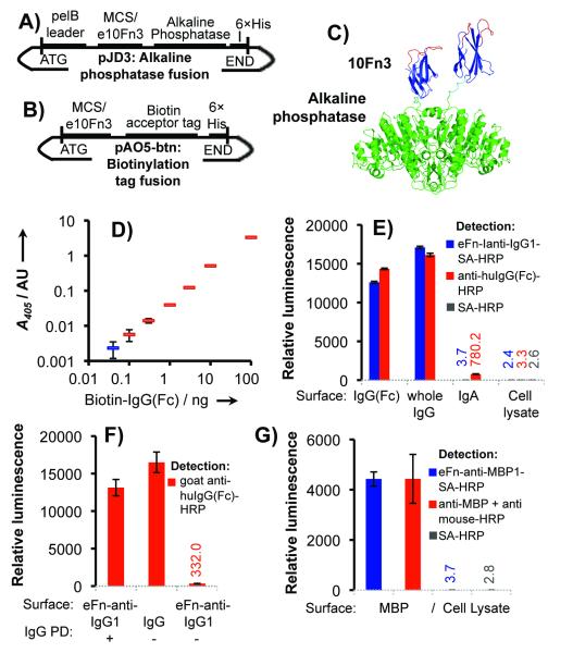
Figure 3.
Validation of binders for use in enzyme-linked detection assays. (a) pJD3 vector for creating e10Fn3-alkaline phosphatase fusions and (b) pAO5-btn vector for generating fusions to a biotin acceptor peptide tag. (c) Structural model of 10Fn3-AP fusions. The 10Fn3 scaffold is blue, randomized loops are red, and alkaline phosphatase is green. 10Fn3 domains (PDBID: 1FNF and 1FNA) are joined to AP (PDBID: 1ALK) by a GSGGSG linker (cyan). (d) Mock sandwich antibody-free ELISA assay for IgG detection. (Blue is the no-IgG control). (e) Comparison of eFn-anti-IgG-SA-HRP to anti human IgG-HRP. (f) eFn-anti-IgG1 efficiently captures IgG (Pull-down, “PD”) for detection by polyclonal anti-huIgG-HRP (g) Comparison of eFn-anti-MBP1-SA-HRP to a commercially available monoclonal. Binding assays were performed in triplicate.

