
Figure 1.
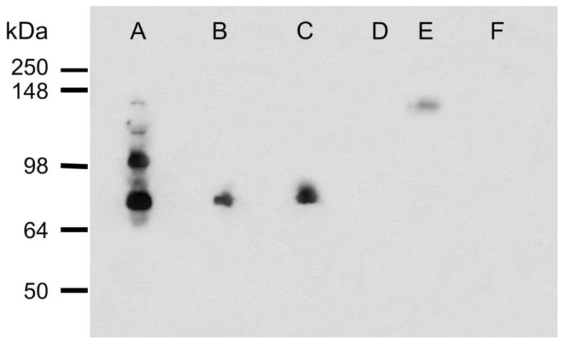
Western blot analysis of fesselin immunoreactivity. Gizzard (A), thigh (B), breast (C) and heart (D, E) are shown. Lane (E) was loaded with 10-times the amount of protein used in other lanes. Pancreas extract (F) was used as a negative control tissue.