
Figure 2.
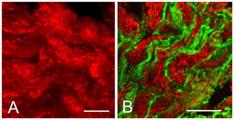
Immunocytochemical localization of fesselin in chicken gizzard. (A) Specific staining was observed primarily as punctuate rod-like intracellular inclusions similar in appearance and distribution to that of cytoplasmic dense bodies. (B) Fesselin (red) did not co-localize with the plasma membrane (dense plaques) associated protein talin (green). Bar = 10 μm.