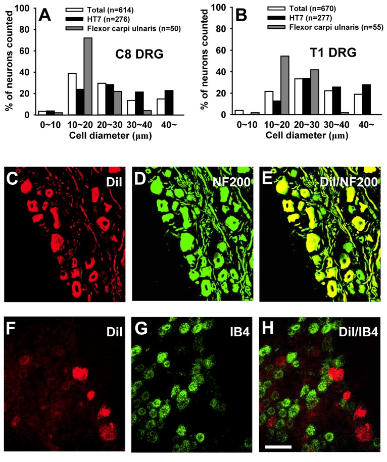
Figure 7. Local afferent fiber composition at HT7 acupoint contains middle- or large-sized A fiber.
A, B: Size distribution of C8- and T1-DRG neurons labeled retrogradely after injection of DiI into HT7 acupoints. ‘Total’ indicates histograms of total DRG cell size distribution. And ‘Flexor carpi ulnaris’ indicates histograms the cell size distribution of labeled DRG neurons after injection of DiI into a muscle near HT7. C, D: Most DiI-labeled DRG neurons are double-stained with A-fiber marker NF200 (C), but not C-fiber marker IB4 (D). Bar=100 μm.

