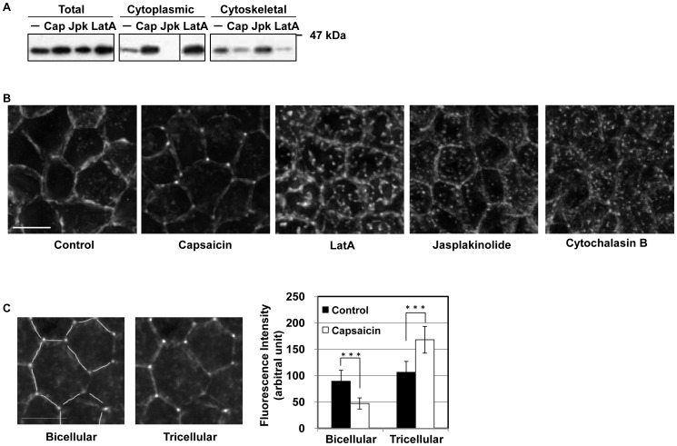
Figure 2. Capsaicin induces actin depolymerization and morphological changes.
(A) Monolayers were exposed to ethanol control, capsaicin (300 µM, 45 min), Jpk (2 µM, 60 min) or LatA (0.5 µM, 90 min) and lysed, and the G- and F-actin fractions were separated. Fractions were analyzed by SDS-PAGE and immunoblotting with an anti-actin antibody. (B) MDCK monolayers were exposed to ethanol control, capsaicin (300 µM, 45 min), LatA (0.1 µM, 30 min), Jpk (2 µM, 1 min) or CytoB (5 µg/ml, 15 min), and were fixed and stained with rhodamine-phalloidin to detect F-actin. Images from each z-section were deconvoluted and overlayed. Bar: 10 µm. (C) The fluorescence intensities of the images in (B) were analyzed. Bar: 10 µm. ***represents p<0.001 by Student’s t test.

