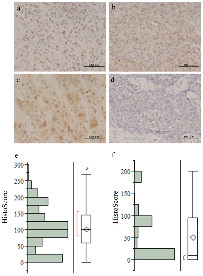
Figure 1. Representative microscopic views of PA1 staining with rabbit polyclonal antibody against PA1.
a, nuclear staining; b, cytoplasmic staining; c, mixed nuclear and cytoplasmic staining; d, negative staining (original magnification × 400) e, histograms for Histo-Score (HS) of PA1-nuc and f, of PA1-cyto (mean; 51.4, S.D.; 62.4). Abbreviations: PA1-nuc, nuclear PA1 expression; PA1-cyto, cytoplasmic PA1 expression; S.D., Standard Deviation.

