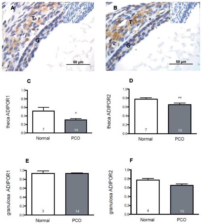
Figure 1. Expression of adiponectin receptors in follicles from normal and polycystic human ovaries.
Both AdipoR1 (Figure 1a) and AdipoR2 (Figure 1b) were detected in granulosa (G) and theca (T) of antral follicles (insets show control [negative] sections). The proportions of theca and granulosa cells expressing protein for adiponectin receptor I & 2 (AdipoR1 and AdipoR2) in follicles from normal and polycystic ovaries are shown in the bar graphs. A total of 920 follicles were analysed. A significant reduction in the proportion of TCs labelled for (c) AdipoR1 and (d) AdipoR2 was demonstrated in polycystic ovaries compared with normal ovaries from healthy women (*p=0.002, **p=0.049, Mann-Whitney). No differences between PCO and control tissue were detected in the proportion of granulosa cells labelling for AdipoR1 (e) or AdipoR2 (f). Data shown are mean + SEM in up to 7 normal and 15 polycystic ovaries.

