
Figure 5. Western blot analysis of cleaved PARP in the lymphocytes treated with roscovitine.
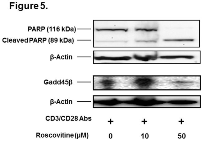
Mouse CD4+ T cells were stimulated with anti-CD3 and anti-CD28 antibodies in the presence of 0, 10, and 50 μM roscovitine. Seventy two hours later, PARP and Gadd45β in the cell lysates were analyzed by western blot. In addition, β-actin was measured as a loading control. Shown is the representative image of western blot analysis from 2 independent studies.