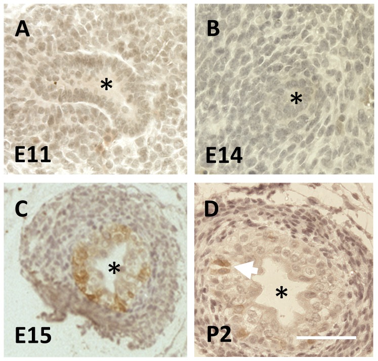
Figure 1. CK15 immunohistochemistry of wild type fetal mouse ureters.
In these brightfield images, positive CK15 immunostaining is brown. Nuclei were counterstained blue with haematoxylin. In each image, the asterisk indicates the lumen. A and B. CK15 was neither detected in the E11 UB (A) nor in the monolayered urothelium of the ureteric stalk at E14 (B). C. A subset of urothelial cells in the multilayered E15 urothelium were positive for CK15. D. At postnatal day two (P2), CK15 was detected in a subset of uroltheial cells (two such adjacent cells are indicated by the white arrow). Bar is 200 μm.

