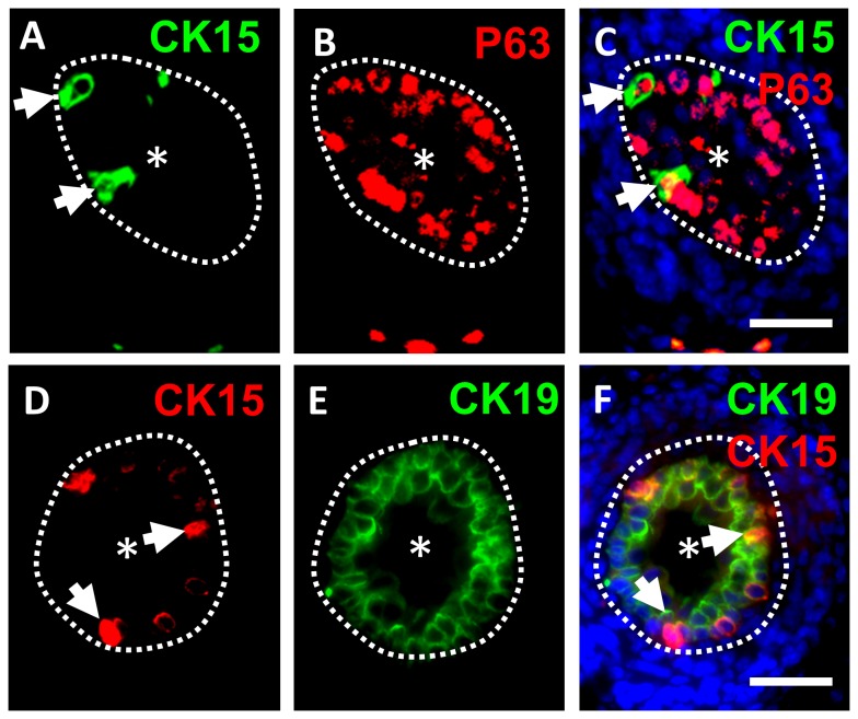
Figure 2. Double immunostaining for CK15/P63 and CK15/CK19.
Fluorescence images of cross sections of wild type E17 mouse ureters. In each frame, the asterisk indicates the ureteric lumen and the dotted lines indicate the border between the urothelium and differentiating smooth muscle. A-C. Double immunostaining for CK15 (green in A) and P63 (red in B), with the merged images shown in C where nuclei are stained blue with DAPI. The same two CK15+ cells are arrowed in A and C; note the presence of P63 in their nuclei. D-F. Double immunostaining for CK15 (red in D) and CK19 (green in E), with the merged images shown in F where nuclei have been stained blue with DAPI. The same two CK15+ cells are arrowed in D and F. Note that CK19 has an overlapping but more extensive distribution than CK15. Bars are 100 μm.

