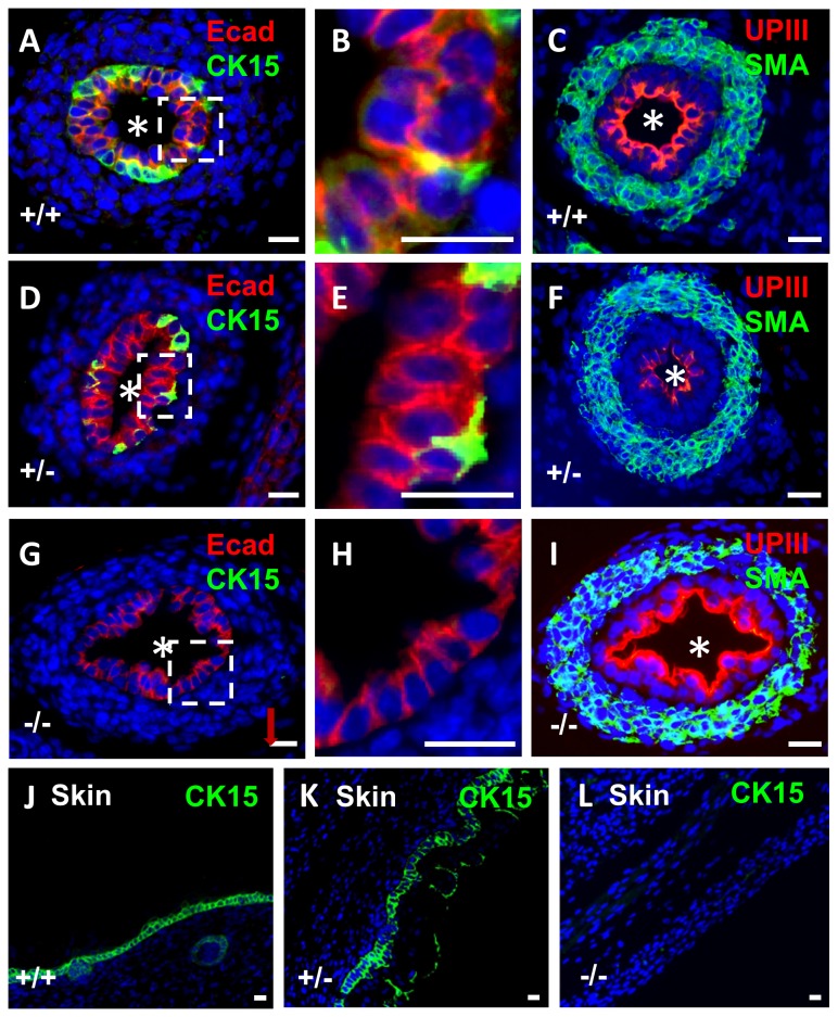
Figure 3. Immunostaining for CK15 in wild type and p63 mutant embryonic mice.
All frames are fluorescence images of E17 organs stained with DAPI (blue nuclei). A-C and J. Tissues from wild type (+/+) mice, D-F and K. Tissues from p63 heterozygous mice. G-I and L. Tissues from homozygous p63 mutant mice (-/-). A-I are ureter cross sections, the asterisks indicating the lumen. The boxed areas around portions of the urothelium in A, D and G have been respectively enlarged and depicted in frames B, E and H to show the layer(s) of cells. J, K and L are sections through the epidermis. In wild type ureters, CK15 was immunodetected in a subset of basal urothelia (green in A and B) whereas E-cadherin immunoreactivity (Ecad; red in A and B) was detected in all cells of the multilayered urothelium. In wild type ureters, uroplakin III (UPIII; red in C) was detected in the most superficial urothelial layer and α-smooth muscle actin (SMA; green in C) was immunodetected in the ureteric wall. In p63 heterozygous mice (D-F), immunolocalisation patterns for CK15, E-cadherin, uroplakin III and α-smooth muscle actin were similar to wild types. The ureteric urothelium of homozygous p63 mutant mice (G) contained abnormal sections with a monolayer of cells (G and H) and CK15 was not detected in these organs (G and H). In contrast to CK15, prominent uroplakin III immunostaining and α-smooth muscle actin were immunodetected in p63 homozygous ureters. CK15 was immonodetected (green) in wild type (J) and p63 heterozygous (K) epidermis. CK15 was not immunodetectable in homozygous p63 mutant epidermis (L). Bars are 20 μm.

