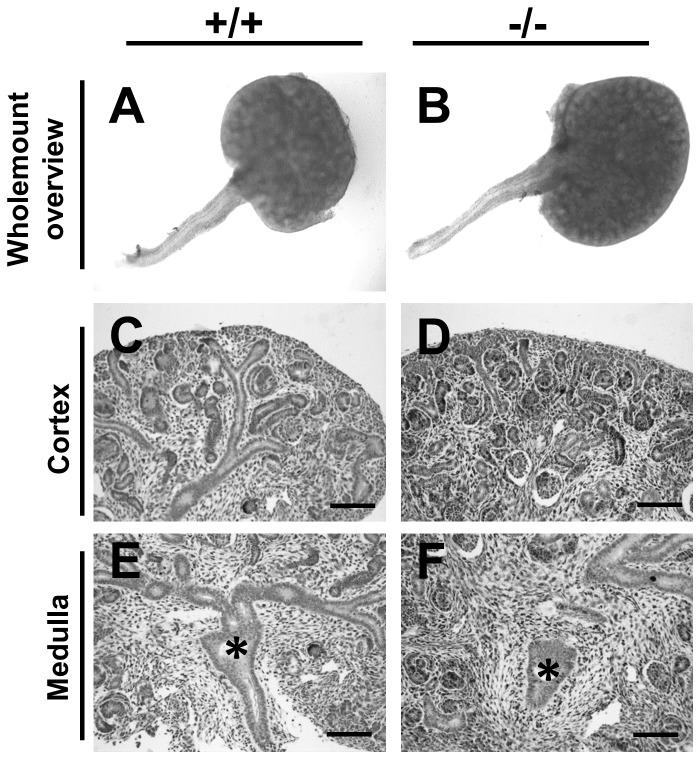
Figure 4. Fetal kidneys and ureters of wild type and p63 mutant mice.
All frames are from E14 organs. A, C and E are from wild type mice (-/-) and B, D and F are from p63 homozygous mutant mice (-/-). A and B. Whole mount images showing similar gross appearance of renal tracts in the two genotypes. C-F. Histology sections of the cortex (C and D) and medulla (E and F) of the metanephric kidney, with nuclei stained with haematoxylin. Both show several layers of forming nephrons in the cortex and a normal-looking nascent kidney pelvis (asterisks) in the medulla. Bars are 100 μm.

