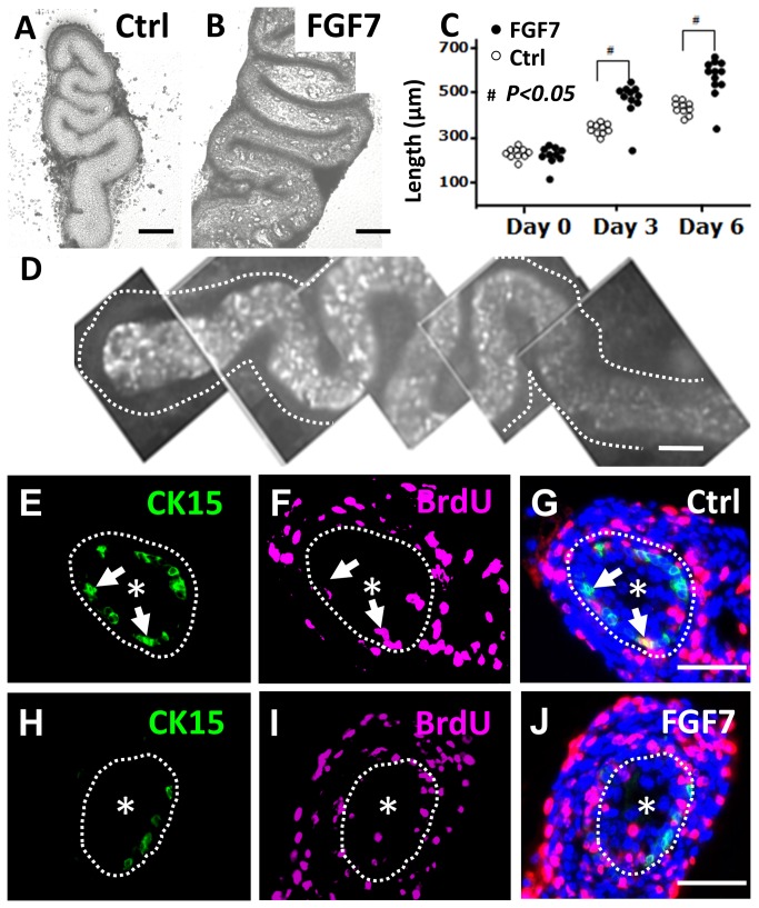
Figure 6. Growth of explanted embryonic ureters.
E14 wild type mouse ureters were explanted into organ culture and maintained for up to six days. A and B. Phase contrast images of whole explants on day six of culture. Ureters cultured in the presence of FG7 (B) appeared bulkier than controls (Ctrl in A). C. Groups of explants cultured with FGF7 were significantly (#; p<0.05, as assessed by t-tests) longer than controls at three and six days of culture (each dot represents one ureter). D. Whole mount CK15 immunostaining of ureteric explant on day six of culture showed positive cells in the central, urothelial, core of the organ; the dotted line indicates the perimeter of the ureter and the top/proximal end is on the left of the frame. E-J. Cross sectional images of explanted ureters fed for six days with control media (E-G) or media supplemented with FGF7 (H-J). In each frame, the dotted line marks the border between the urothelium and the surrounding differentiating smooth muscle, and an asterisk has been placed in the lumen. Images E and H demonstrate that CK15+ (green) urothelial cells were present in both experimental groups. Images F and I show BrdU+ cell nuclei in each experimental group. Most of them were in the developing muscle layer but some were present in the urothelium. In the merged images (G and J, where DAPI/blue nuclear staining is also shown), it is apparent the the BrdU+ cells in the urothelium usually do not correspond to the CK15+ cells. In each of the frames E-G, the locations of the same two CK15+ cells are arrowed; the upper cell is BrdU- while the lower one is BrdU+. In H-J, there is a cluster of BrdU+ epithelial nuclei at ‘6-9 o’clock’ and they are separate from CK15+ cells. Bars are 50 μm.

