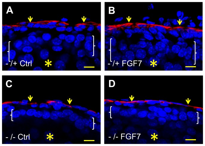
Figure 7. Fluorescence confocal images of embryonic ureter explants.
E14 ureters were explanted and maintained for six days in organ culture, without (Ctrl) or with addition of FGF7 (FGF7). In these longidutinal sections, all nuclei were stained blue with DAPI and the red color indicates positive signal after immunostaining for α-smooth muscle actin, thus marking the muscle layer (small arrows). In each frame the asterisk indicates the (collapsed) lumen and the white brackets span the epithelial layer. Note that the heterozygous p63 explant displays the expected normal epithelium with 2-4 cell layers (A) and that the epithelium is also multilayerd upon exposure to FGF7 (B). In contrast, the urothelium in homozygous mutant p63 (-/-) explants generally have only one or two layers, both without (C) or with (D) FGF7 treatment. Bars are 10 μm.

