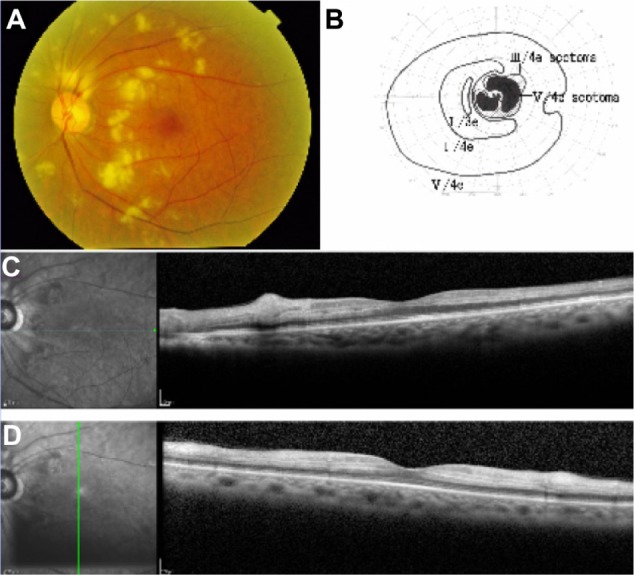
Figure 1.

Clinical findings at presentation.
Notes: (A) A color photograph of the fundus of the left eye shows diffuse retinal whitening. (B) Goldmann kinetic perimetry of the left eye shows paracentral scotomas. (C and D) Optical coherence tomography of the left eye shows edema in the inner retinal layers.
