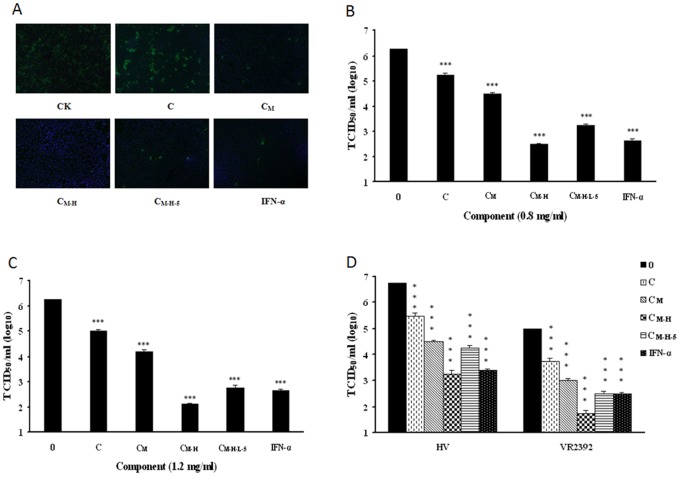
Figure 5. Different C. volvatus fractions blocked PRRSV replication in Marc-145 cells and PAMs.
(A, B and C) Marc-145 cells were infected with PRRSV Ch1a at an MOI of 0.1, and then treated with different C. volvatus extracts at a concentration of 0.8 mg/ml or INF-α (10 units/µl). At 24 h post-infection, cells were fixed and analysed by IFA using antibody against PRRSV N protein (A). The abbreviations at the bottom of fluorescent pictures are explained as follows: C: Aqueous extract of C. volvatus; CM: C was filtered through a Millipore ultrafiltration membrane and the ultrafiltrate gave the resulting crude extract CM; CM-H: CM absorbed on HP-2MGL was eluted with 30% ethanol, which resulted in CM-H; CM-H-L-5: CM-H was loaded on a DEAE and Sephadex LH-20 chromatography column sequentially, which resulted in CM-H-L-5. Virus yield in the supernatant was quantified (B). The same experiment was carried out with different C. volvatus extracts at a concentration of 1.2 mg/ml or INF-α (10 units/µl) (C). (D) Different C. volvatus fractions inhibited replication of VR2332 and HV strains in PAMs at a concentration of 1.2 mg/ml or INF-α (10 units/µl). Virus yield in the supernatant was also quantified. Culture treated with normal saline was set up as negative control (0 mg/ml), and that treated with INF-α was set up as positive control. The brightness of the fluorescence represents the level of the virus. Data are representative of three independent experiments (mean ± SD). Statistical significance was analyzed by Student’s t test. *P<0.05; **P<0.01; ***P<0.001.

