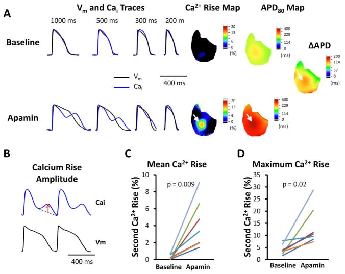
Figure 4.
Secondary rises of Cai in failing ventricles. A. Representative Vm, Cai traces, secondary rises of calcium and APD maps at different PCLs without and with 100 nmol/l apamin perfusion. Apamin prolonged APD, along with secondary rises of Cai, especially at longer PCLs. The areas with the secondary rises of Cai co-localized with areas with the most significant APD prolongation (white arrows). The delta APD indicates the difference of APD after and before apamin. B. The amplitude of secondary Cai rises is defined as the largest deviation from a line drawn between the onset and offset of the secondary Cai rises, as indicated by the red arrow. C. Comparison of average secondary rises of Cai amplitude among all available ventricular pixels at baseline and in the presence of 100 nmol/l apamin of all hearts studied. D. Comparison of maximal amplitude of secondary rises of Cai in each heart at baseline and in the presence of apamin of all hearts studied.

