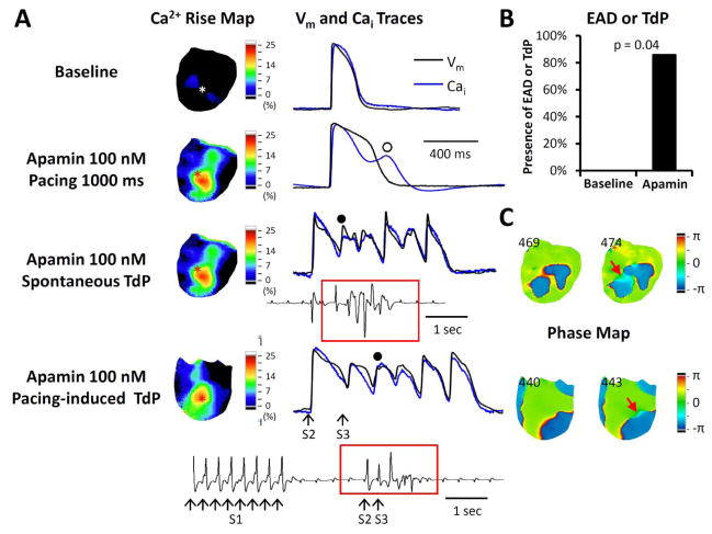
Figure 5.
Relationship between secondary rises of Cai and the development of EAD and PVBs in failing hearts with AV block. A. The Vm and Cai traces were taken from the site marked by an asterisk in the Cai rise map (map of secondary rise of Cai). In this pixel, the Cai tracing tracked the Vm tracing at baseline. Apamin prolonged APD and induced secondary rise of Cai (unfilled circle). Subsequently, spontaneous TdP developed from the same site with a secondary rise of Cai. In addition to spontaneous TdP, short-long-short (30 short S1S1 beats, a long S1S2 and a short S2S3 intervals) pacing protocol also induced TdP in this ventricle, as shown in the bottom tracing. B. Apamin increased EADs and/or TdP inducibility in failing hearts. None of the hearts had EADs at baseline and 6 of 7 hearts developed EADs during apamin perfusion. C. Phase maps of corresponding TdP beats (filled circles in panel A) in the spontaneous and the pacing-induced TdPs. Red arrows point to an area with light blue color (phase change), which is the earliest activation sites of the TdP beats. The numbers indicate frame.

