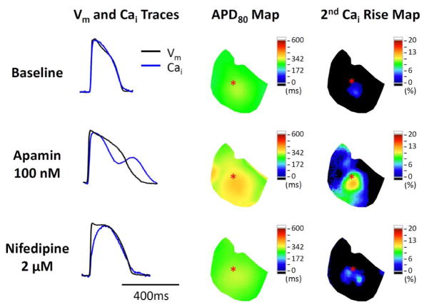
Figure 7.
Representative traces and maps in a failing heart at baseline, during apamin perfusion, and after adding nifedipine. Nifedipine shortened APD and ameliorated secondary rises of Cai. The Vm and Cai tracings were obtained from the site labeled by an asterisk on the APD80 map and secondary Cai rise map. Note that the latter two maps show the co-localization of the secondary rises of Cai and the prolonged APD80 in the same ventricle.

