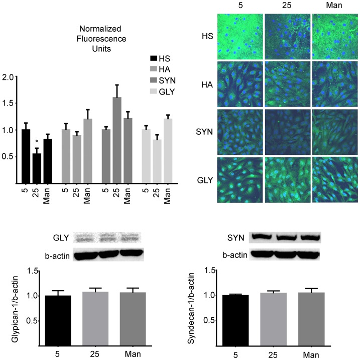
Figure 2. Glycocalyx of BAEC monolayers cultured in 5.
BAEC monolayers were cultured in either normal (5 mM), high (25 mM) glucose or Mannitol control (25 mM) media. After six days, they were immuno-stained with antibodies against Heparan Sulfate (HS), hyaluronic acid (HA), syndecan-1 (SYN) and glypican-1 (GLY) as described in the methods. The monolayers were observed under a confocal microscope and four random fields were chosen for each condition. Only the mean fluorescence representative of Heparan Sulfate was significantly decreased to 55.5±11% in HG (p = 0.023). In addition, the contents of the core proteins Syndecan-1 and Glypican-1 were determined by western blot and consistenly showed that neither high glucose nor Mannitol had an effect on these components. (p-value >0.05, n = 4).

