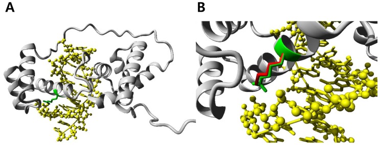
Figure 2. Molecular modeling of the POU4F3 protein constructed by the 2xsd protein as the template.

(A) Overview of the modeled POU4F3 domain. The protein chain is shown in grey using a ribbon representation, the position of the mutated residue is indicated in green, the DNA is shown as a ball-and-stick model and colored yellow. (B) A close-up view of the mutated residue. The side chains of the wild-type and the mutant residue are shown in green and red, respectively. The figures show that the wild-type Arginine residue interacts with the phosphate backbone of the DNA, while the mutant Lysine residue is slightly shorter than Arginine and cannot make the same interactions with DNA.
