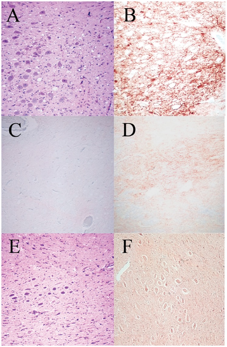
Figure 1. Central Nervous system pathology.
Examples of vacuolation and immunostaining for PrPSc in brain tissue. A. Haematoxylin and Eosin (H&E) staining showing vacuolation in the dorsal motor vagus nucleus of L55 (VRQ/VRQ) had natural scrapie-like disease (x100). B. Strong immunostaining for PrPSc (diagnostic for natural scrapie) with BG4 antibody in the dorsal motor vagus nucleus of L55 (x40). C. Marginal immunostaining for PrPSc (diagnostic for SSBP/1) with BG4 in the dorsal motor vagus in the brain of L111 (VRQ/ARR) (x 100). D. Marginal immunostaining for PrPSc (diagnostic for SSBP/1) with BG4 in the dorsal motor vagus of L101 (VRQ/VRQ) (x100). E. Nil vacuolation (typical for SSBP/1) in an H&E section of the dorsal motor vagus in the brain of L101 (VRQ/VRQ) (x100). F. Marginal immunostaining for PrPSc (diagnostic for SSBP/1) in the brain of L106 (VRQ/VRQ) (x100).

