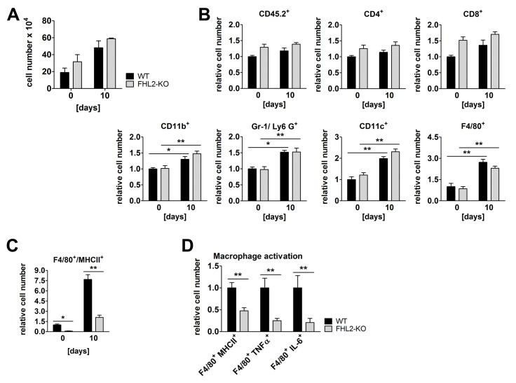
Figure 4. Immune status of WT and FHL2-KO lungs.
(A) Cell count in BALF of control and 10-day BLM-treated mice. N = 4 and 2 for control and BLM-treated mice, respectively, with 3 to 4 mice per group in each experiment. (B) Single cell suspensions of lung tissue from control or 10-day BLM-treated mice were studied for the indicated surface receptors by flow cytometry. A total of 105 cells per lung were analysed. n = 8 and 9 for control and n = 5 and 6 for BLM-treated WT and FHL2-KO mice, respectively. The relative mean amount of immune cells presented in the lungs of wild type mice was always assigned a value of 1. (C) Lung cell suspensions from (B) were analysed for MHCII and F4/80 surface marker. (D) The F4/80-positive macrophages of BLM-induced mice were further analysed for expression of activation markers: the MHCII receptor and the intracellular TNFα and IL-6 proteins.

