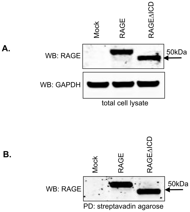
Figure 2. RAGEΔICD is expressed in cells and is present on the cell surface.
A–B. Cell surface biotinylation was performed with C6 glioma cells expressing RAGE, RAGEΔICD, or empty vector (mock). Western blot for RAGE using polyclonal anti-RAGE antibodies was performed using total input of cell lysate (A) and extracts subjected to cell surface biotinylation which was followed by pull-down with streptavidin agarose (B). GAPDH was detected in total cell lysate by western blot using monoclonal anti-GAPDH for normalization.

