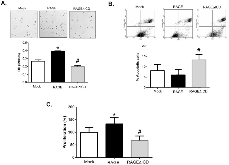
Figure 6. RAGEΔICD alters cell adhesion and viability.
A. Cell adhesion assays were performed by seeding C6 glioma cells expressing RAGE, RAGEΔICD, or empty vector (mock) onto cell culture dishes for 2 hours. Adhered cells were fixed and stained with crystal violet before quantification by a spectrophotometer. B. Apoptosis was assessed by staining C6 glioma cells expressing RAGE, RAGEΔICD, or empty vector (mock) for Annexin V (X-axis) and PI (Y-axis) which as followed by flow cytometric analysis. C. Proliferation was assessed using the Alamar Blue reagent and assessed after 48 hours of culture of C6 glioma cells expressing RAGE, RAGEΔICD, or empty vector (mock). Data are means ± SEM from three independent experiments. *, significant differences (P≤0.05) between mock and RAGE, whereas # significant differences (P≤0.05) between RAGE and RAGEΔICD.

