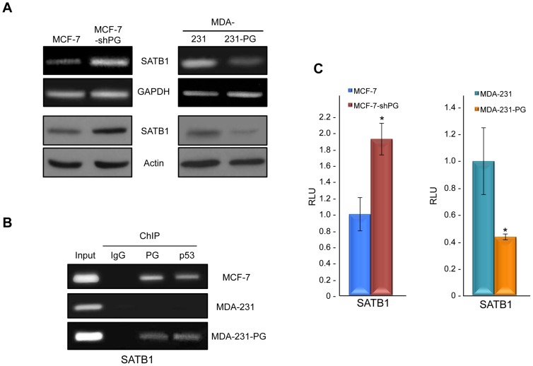
Figure 2. Plakoglobin suppresses SATB1 in mammary epithelial cell lines.
A. (Top) Total cellular RNA was isolated from MCF-7, MCF-7-shPG, MDA-231 and MDA-231-PG cells, reverse transcribed and processed for PCR using primers specific to SATB1 and GAPDH. (Bottom) Equal amounts of total cellular proteins from these cells were resolved by SDS-PAGE and processed for immunoblotting with antibodies to SATB1 and Actin.B. MCF-7, MDA-231 and MDA-231-PG cells were formaldehyde fixed and processed for chromatin immunoprecipitation as described in Fig. 1B. The purified DNA was then processed for PCR using SATB1 primers. As a positive control, total cellular DNA (Input) was amplified using the same primers. C. MCF-7, MCF-7-shPG, MDA-231 and MDA-231-PG cells were transfected with luciferase reporter constructs and processed as described in Fig. 1C. The SATB1 promoter activity was normalized to the corresponding vector activity for each cell line and then normalized to MDA-231 or MCF-7, respectively (*p<0.01). PG, plakoglobin; RLU, Relative Light Units.

