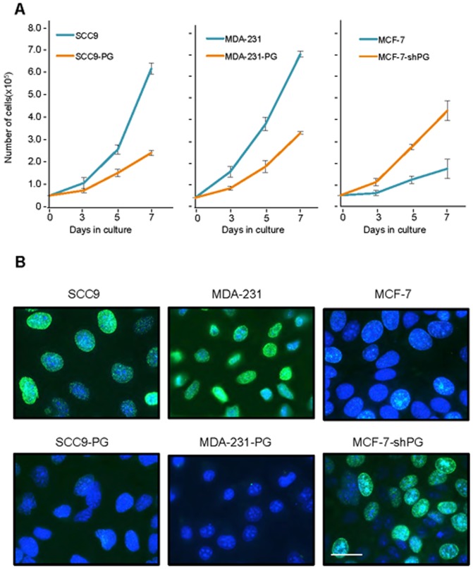
Figure 5. Plakoglobin decreases in vitro cell growth and proliferation.
A. Replicate cultures of SCC9, SCC9-PG, MDA-231, -231-PG, MCF-7 and MCF-7-shPG cells were established at single cell density and cells were counted at 3, 5 and 7 days. Each time point represents the average of three independent experiments. The absence of error bars at some time points is due to the small differences among the experiments. B. SCC9, SCC9-PG, MDA-231, -231-PG, MCF-7 and MCF-7-shPG cells were plated on glass coverslips and allowed to grow for 6 days at which time BrdU was added to the cell cultures for 24 hours. BrdU incorporation was then assessed by immunofluorescence staining using BrdU antibodies. Nuclei were countersatined with DRAQ5 and cells viewed using a 63X objective of an LSM510 META (Zeiss) laser scanning confocal microscope. Bar, 20 μm.

