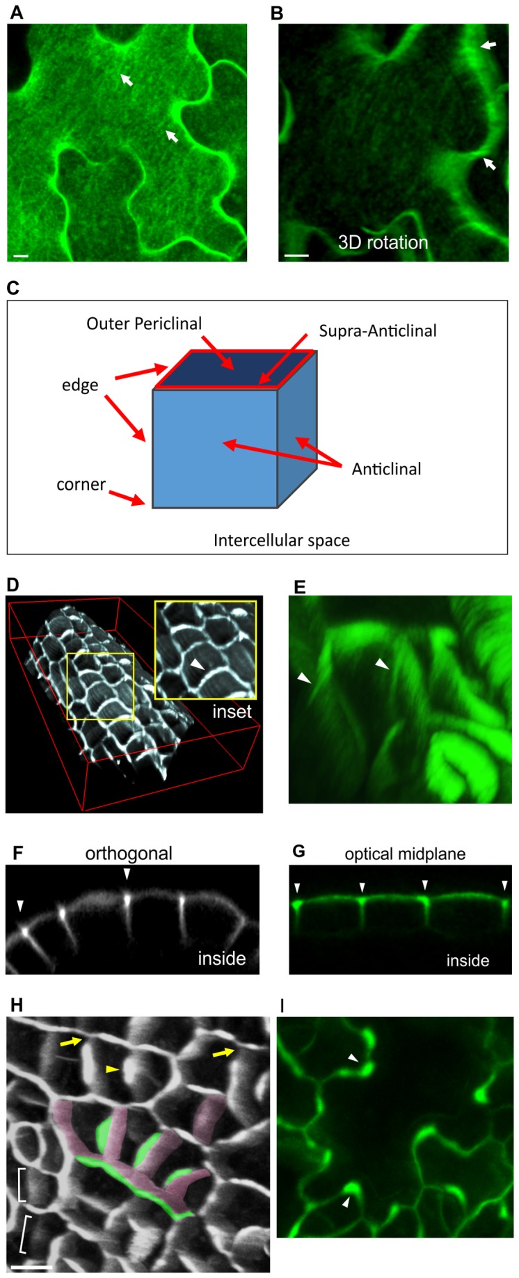
Figure 1. YFP-LTPG shows filamentous patterning and accumulation over anticlinal walls.

A Mature cotyledon epidermal cell showing filamentous YFP-LTPG patterning (arrows).
B 3D rotation of mature cotyledon epidermal cell shows filaments extending down anticlinal walls (arrows).
C Schematic diagram showing nomenclature used in this article.
D Tilted image from z-series 3D reconstruction showing anticlinal YFP-LTPG accumulation in unexpanded leaf petiole cells (arrowheads).
E Radial striations (arrowheads) along anticlinal walls of unexpanded leaf petiole cells. Tilted image from 3D rotation.
F Orthogonal slice from confocal Z-series illustrates YFP-LTPG fluorescence accumulation over anticlinal walls (arrowheads).
G Optical midplane image of epidermal cells showing accumulation of YFP-LTPG over anticlinal walls (arrowheads).
H Tilted image from 3D reconstruction of YFP-LTPG expressed in unexpanded leaf. Recently-formed cell walls (brackets) contain faint, homogeneous fluorescence. As the anticlinal wall matures, YFP-LTPG accumulates above it non-uniformly (green highlight), showing a gradual increase in fluorescence with increasing distance from three-way junctions (arrows). Green highlight shows outer edge enrichment; pink shows anticlinal walls. Arrowheads indicate example of outer enrichment site. Bottom panel shows orthogonal slice.
I Mid-stage lobed leaf epidermal cell showing non-uniform accumulation of YFP-LTPG over anticlinal walls. Arrowheads indicate accumulation of YFP-LTPG within concave sides of cell lobes. Bars, 10 µm.
