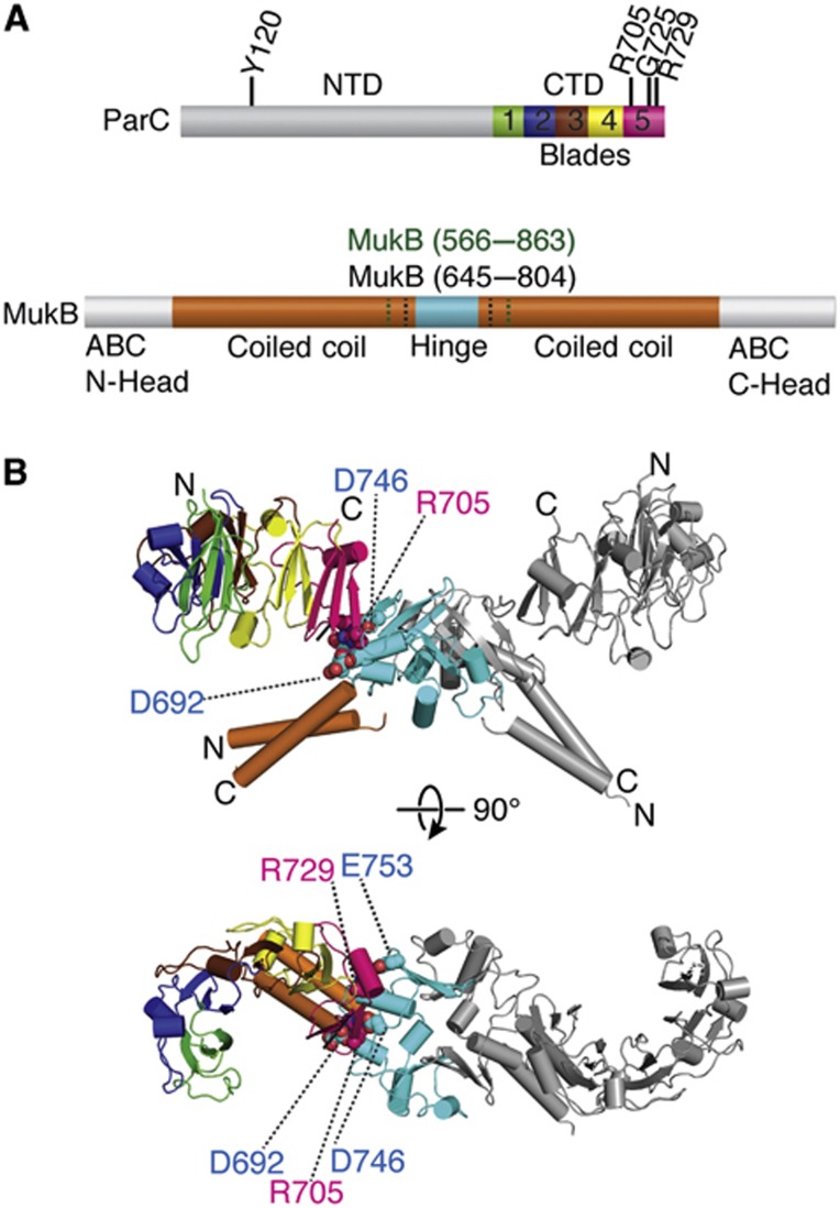
Figure 1.
Structure of a MukB hinge·topo IV complex. (A) Primary structure and domain organization of E. coli ParC and MukB. A subset of the ParC residues addressed in this study are labelled on the primary structure. MukB hinge constructs (aa 566–863 (green), aa 645–804 (black)) used here are also denoted. Different subdomain repeats of the CTD (termed as ‘blades’) are coloured rainbow. (B) Crystal structure of the hinge CTD heterotetramer in cartoon representation. Colouring for one MukB hinge and ParC CTD is as per (A), with dimer-related protomer coloured grey. Residues mutated and assayed in this study are depicted as spheres and labelled.

