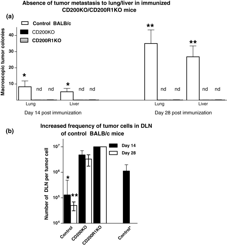
Fig. 1.
Comparison of lung and liver metastases (a) and frequency of tumor cells cloned from DLN (b) in control, CD200KO or CD200R1KO BALB/c mice receiving 5 × 10⁵ EMT6 tumor cells subcutaneously into the mammary fat pads, followed by surgical resection 15 days later, and immunization with EMT6 with CpG as adjuvant. Eight mice were used per group, with four of each sacrificed at 14/28 days post-surgery to measure macroscopic tumor metastases in the lung/liver (a). DLN cells harvested from individual mice were cultured under limiting dilution for 3 weeks to assess the frequency of tumor cells cloned (b). All data represent arithmetic means (±SD) for each group. Data to right in b (control *) indicates frequency of tumor cells in DLN of an independent group of mice at day 15 (the day of surgical resection). nd in a indicates no metastases detected; *,**p < 0.05 relative other groups at day 14/28, respectively

