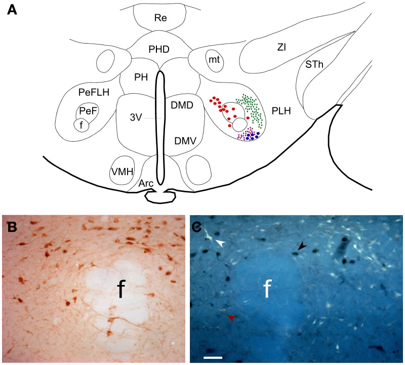
Figure 2.
(A) Schematic drawing of the hypothalamic area in a coronal section of rat brain at antero-posterior coordinates Bregma −3.36 mm. It is shown the distribution of neurons projecting to PnO (red dots) and LC (blue dots) and the distribution of fiber terminals arising PnO and LC are represented in green and purple small dots, respectively. (B) Microphotograph of PeF showing neurons immunocytochemically stained for anti-Orx. (C) Microphotograph showing adjacent section showed in (B) where Orx neurons are pointed by black arrowheads, FG neurons projecting to LC by white arrowheads and double labeled neurons by red arrowheads. Calibration bar: 50 μm. Abbreviations: 3V: 3rd ventricle; Arc: arcuate hypothalamic nucleus; DMD: dorsomedial hypothalamic nucleus, dorsal part; DMV: dorsomedial hypothalamic nucleus, ventral part; f: fornix; mt: mammillothalamic tract; PeF: perifornical nucleus; PeFLH: perifornical part of lateral hypothalamus area; PH: posterior hypothalamic nucleus; PHD: posterior hypothalamic area, dorsal part; PLH: peduncular part of lateral hypothalamus; Re: reuniens thalamic nucleus; Sth: subthalamic nucleus; VMH: ventromedial hypothalamic nucleus; ZI: zona incerta.

