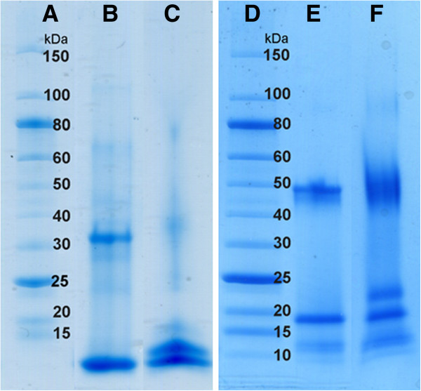
Figure 3.

SDS-PAGE analysis of the PEGylation products. Left, the P6c peptide: A) Mw Marker B) uncoupled C) coupled with PEG; Right, the P6c-PbK peptide: D) Mw Marker E) uncoupled; F) coupled with PEG. For both constructs also the trimer band is visible on the gel.
