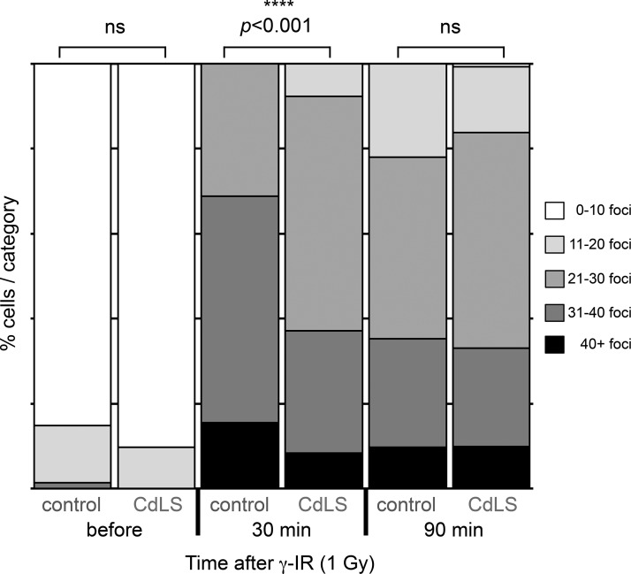
Figure 4.
53BP1 foci formation is reduced at an early time point in NIPBL-deficient LCLs. LCLs from healthy controls or LCLs deficient in NIPBL (P1-P3, P5) were exposed to γ-IR (1Gy) and harvested, cytospun, and fixed at the indicated time points. The cells were then stained with anti-53BP1 antibodies and 53BP1 foci were detected using a confocal microscope. Image stacks were exported to ImageJ and 53BP1 foci were quantified in ∼40 nuclei per LCL per time point in each experiment. The bars represent the relative distribution of 53BP1 foci at indicated time points before and after γ-IR. Two independent experiments were performed. Statistically significant differences are highlighted with stars (Mann-Whitney U test).

